Overview Of Hydatidiform Mole
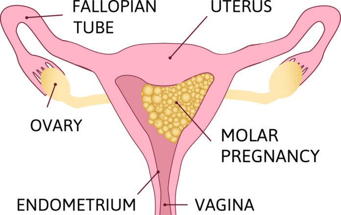
Hydatidiform Mole (HM) is a rare mass or growth that forms inside the womb (uterus) at the beginning of a pregnancy. It is a type of gestational trophoblastic disease (GTD).
Commonly Associated With
Hydatid mole; Molar pregnancy; Hyperemesis – molar
Causes Of Hydatidiform Mole
HM, or molar pregnancy results from abnormal fertilization of the oocyte (egg). It results in an abnormal fetus. The placenta grows normally with little or no growth of the fetal tissue. The placental tissue forms a mass in the uterus. On ultrasound, this mass often has a grape-like appearance, as it contains many small cysts.
The chance of mole formation is higher in older women. A history of mole in earlier years is also a risk factor.
A molar pregnancy can be of two types:
Partial molar pregnancy: There is an abnormal placenta and some fetal development.
Complete molar pregnancy: There is an abnormal placenta and no fetus.
There is no way to prevent the formation of these masses.
Symptoms Of Hydatidiform Mole
Symptoms of molar pregnancy may include:
Abnormal growth of the uterus, either bigger or smaller than usual
Severe nausea and vomiting
Vaginal bleeding during the first 3 months of pregnancy
Symptoms of hyperthyroidism, including heat intolerance, loose stools, rapid heart rate, restlessness or nervousness, warm and moist skin, trembling hands, or unexplained weight loss
Symptoms similar to preeclampsia that occur in the first trimester or early second trimester, including high blood pressure and swelling in the feet, ankles, and legs (this is almost always a sign of a hydatidiform mole because preeclampsia is extremely rare this early in a normal pregnancy)
Exams & Tests
Your health care provider will perform a pelvic exam, which may show signs similar to a normal pregnancy. However, the size of the womb may be abnormal and there may be no heart sounds from the baby. Also, there may be some vaginal bleeding.
A pregnancy ultrasound will show a snowstorm appearance with an abnormal placenta, with or without some development of a baby.
Tests done may include:
hCG (quantitative levels) blood test
Abdominal or vaginal ultrasound of the pelvis
Chest x-ray
CT or MRI of the abdomen (imaging tests)
Complete blood count (CBC)
Blood clotting tests
Kidney and liver function tests
Treatment Of Hydatidiform Mole
If your provider suspects a molar pregnancy, removal of the abnormal tissue with a dilation and curettage (D&C) will most likely be suggested. D&C may also be done using suction. This is called suction aspiration (The method uses a suction cup to remove contents from the uterus).
Sometimes a partial molar pregnancy can continue. A woman may choose to continue her pregnancy in the hope of having a successful birth and delivery. However, these are very high-risk pregnancies. Risks may include bleeding, problems with blood pressure, and premature delivery (having the baby before it is fully developed). In rare cases, the fetus is genetically normal. Women need to completely discuss the risks with their providers before continuing the pregnancy.
A hysterectomy (surgery to remove the uterus) may be an option for older women who DO NOT wish to become pregnant in the future.
After treatment, your hCG level will be followed. It is important to avoid another pregnancy and to use a reliable contraceptive for 6 to 12 months after treatment for a molar pregnancy. This time allows for accurate testing to be sure that the abnormal tissue does not grow back. Women who get pregnant too soon after a molar pregnancy is at high risk of having another molar pregnancy.


