Overview
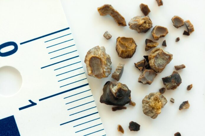
A kidney stone is a hard object that is made from chemicals in the urine. There are four types of kidney stones: calcium oxalate, uric acid, struvite, and cystine. A kidney stone may be treated with shockwave lithotripsy, uteroscopy, percutaneous nephrolithomy, or nephrolithotripsy. Common symptoms include severe pain in the lower back, blood in the urine, nausea, vomiting, fever, and chills, or urine that smells bad or looks cloudy.
Urine has various wastes dissolved in it. When there is too much waste in too little liquid, crystals begin to form. The crystals attract other elements and join together to form a solid that will get larger unless it is passed out of the body with the urine. Usually, these chemicals are eliminated in the urine by the body’s master chemist: the kidney. In most people, having enough liquid washes them out or other chemicals in urine stop a stone from forming. The stone-forming chemicals are calcium, oxalate, urate, cystine, xanthine, and phosphate.
After it is formed, the stone may stay in the kidney or travel down the urinary tract into the ureter. Sometimes, tiny stones move out of the body in the urine without causing too much pain. But stones that don’t move may cause a back-up of urine in the kidney, ureter, bladder, or urethra. This is what causes the pain.
Possible causes include drinking too little water, exercise (too much or too little), obesity, weight loss surgery, or eating food with too much salt or sugar. Infections and family history might be important in some people. Eating too much fructose correlates with an increased risk of developing a kidney stone. Fructose can be found in table sugar and high fructose corn syrup.
Commonly Associated With
Renal calculi; Nephrolithiasis; Stones – kidney; Calcium oxalate – stones; Cystine – stones; Struvite – stones; Uric acid – stones; Urinary lithiasis
Cause
Kidney stones are common. Some types run in families. They often occur in premature infants.
There are different types of kidney stones. The cause of the problem depends on the type of stone.
Stones can form when urine contains too many of certain substances that form crystals. These crystals can develop into stones over weeks or months.
Calcium stones are most common. They are most likely to occur in men between ages 20 to 30. Calcium can combine with other substances to form the stone.
Calcium stones can also form from combining with phosphate or carbonate.
The biggest risk factor for kidney stones is not drinking enough fluids. Kidney stones are more likely to occur if you make less than 1 liter (32 ounces) of urine a day
Kidney stones are caused by high levels of calcium, oxalate, and phosphorus in the urine. These minerals are normally found in urine and do not cause problems at low levels.
Certain foods may increase the chances of having a kidney stone in people who are more likely to develop them.
Symptoms
You may not have symptoms until the stones move down the tubes (ureters) through which urine empties into your bladder. When this happens, the stones can block the flow of urine out of the kidneys.
The main symptom is severe pain that starts and stops suddenly:
• Pain may be felt in the belly area or side of the back.
• Pain may move to the groin area (groin pain), testicles (testicle pain) in men, and labia (vaginal pain) in women.
Other symptoms can include:
• Abnormal urine color
• Blood in the urine
• Chills
• Fever
• Nausea and vomiting
Treatment
Wait for the stone to pass by itself.
Often you can simply wait for the stone to pass. Smaller stones are more likely than larger stones to pass on their own.
Waiting up to six weeks for the stone to pass is safe as long as the pain is bearable, there are no signs of infection, the kidney is not fully blocked and the stone is small enough that it is likely to pass. While waiting for the stone to pass, you should drink normal amounts of water.
You may need medication when there is a lot of pain
Medication
Certain medications have been shown to improve the chance that a stone will pass. The most common medication prescribed for this reason is tamsulosin. Tamsulosin (Flomax) relaxes the ureter, making it simpler for the stone to pass. You may also need pain and anti-nausea medicine as you wait to pass the stone.
Surgery
Surgery may be needed to remove a stone from the ureter or kidney if:
● The stone fails to pass.
● The pain is too great to wait for the stone to pass.
● The stone is affecting kidney work. Small stones in the kidney may be left alone if they are not causing pain or infection. Some people choose to have their small stones removed. They do this because they are afraid the stone will start to pass and cause pain without warning.
Kidney stones should be removed by surgery if they cause repeated infections in the urine or because they are blocking the flow of urine from the kidney. Today, surgery often involves small or no incisions (cuts), minor pain, and minimal time off work.
Other
Common types of Kidney Stones
There are four main types of stones:
1. Calcium oxalate: The most common type of kidney stone is created when calcium combines with oxalate in the urine. Inadequate calcium and fluid intake, as well as other conditions, may contribute to their formation.
2. Uric acid: This is another common type of kidney stone. Foods such as organ meats and shellfish have high concentrations of a natural chemical compound known as purines. High purine intake leads to a higher production of monosodium urate, which, under the right conditions, may form stones in the kidneys. The formation of these types of stones tends to run in families.
3. Struvite: These stones are less common and are caused by infections in the upper urinary tract.
4. Cystine: These stones are rare and tend to run in families.
Exams and Tests
The health care provider will perform a physical exam. The belly area (abdomen) or back might feel sore.
Tests that may be done include:
• Blood tests to check calcium, phosphorus, uric acid, and electrolyte levels
• Kidney function tests
• Urinalysis to see crystals and look for red blood cells in urine
• Examination of the stone to determine the type
Stones or a blockage can be seen on:
• Abdominal CT scan
• Abdominal x-rays
• Intravenous pyelogram (IVP)
• Kidney ultrasound
• Retrograde pyelogram


