Overview
Paget’s disease of the breast is a rare form of breast cancer that almost exclusively occurs in women. However, rare cases have been reported in men. Paget’s disease of the breast is characterized by inflammatory, “eczema-like” changes of the nipple that may extend to involve the areola, which is the circular, darkened (pigmented) region of skin surrounding the nipple. Initial findings often include itching (pruritus), scaling, and crusting of and/or discharge from the nipple. In individuals with Paget’s disease of the breast, distinctive tumor cells (known as Paget cells) are present within the outermost layer of skin (epidermis) of the nipple, when viewed under a microscope. Most women with this disorder have underlying cancer (malignancy) affecting the milk ducts (ductal carcinoma). The milk ducts are the channels that carry milk secreted by the lobes of the breast to the nipple. The exact cause of Paget’s disease of the breast is not fully understood.
Commonly Associated With
- Paget’s Disease Of The Nipple
- Paget’s Disease Of The Nipple And Areola
- Mammary Paget’s Disease
Cause
Doctors do not fully understand what causes Paget’s disease of the breast. The most widely accepted theory is that cancer cells from a tumor inside the breast travel through the milk ducts to the nipple and areola. This would explain why Paget disease of the breast and tumors inside the same breast are almost always found together.
A second theory is that cells in the nipple or areola become cancerous on their own (1, 3). This would explain why a few people develop Paget disease of the breast without having a tumor inside the same breast. Moreover, it may be possible for Paget disease of the breast and tumors inside the same breast to develop independently.
Symptoms
Paget’s disease of the breast is a malignant (cancerous) condition that initially appears as chronic, inflammatory, “eczema-like” changes of the nipple and adjacent areas.
In individuals with Paget’s disease of the breast, initial characteristic skin changes may include the appearance of reddish (erythematous), scaling, crusting, and/or abnormally thickened skin patches (plaques) or lesions on the nipple that may extend to adjacent areas of the areola. Some affected individuals may also have abnormal discharge from the nipple. Additional symptoms may include itching (pruritus) or burning sensations and/or oozing or bleeding of the affected area. Eventually, pain and sensitivity of the affected area may be present. Early on, the skin symptoms of Paget’s disease of the breast may fluctuate, improving only to worsen again. Paget’s disease of the breast usually affects one breast (unilateral), but there are rare cases in which both breasts are involved (bilateral).
The initial skin changes of Paget’s disease of the breast may appear relatively benign and many individuals may overlook such symptoms, mistakenly attributing them to an inflammatory skin condition or infection. As a result, diagnosis may be delayed, often up to six months or more. Most individuals with the condition eventually seek medical attention due to associated itching or burning sensations, soreness, or pain of the affected area.
Most women with Paget’s disease of the breast have an underlying malignancy, which may be completely contained within the milk ducts (ductal carcinoma in situ) or may have invaded the surrounding tissue, potentially spreading to the lymph nodes under the arms (axillary lymph nodes) and other regions of the body (metastatic disease).
Approximately 50 percent or more of affected individuals may have a lump or mass that may be felt (palpated) below the nipple. Some individuals with Paget’s disease of the breast may have additional symptoms or physical findings. For example, in some instances, the nipple may turn inward (retracted nipple).
The overall disease course may vary greatly from one person to another, depending upon the nature and size of an underlying malignancy, whether a palpable breast tumor is present upon diagnosis, whether metastatic disease is present, specific treatment measures followed, and other possible factors.
Treatment
Treatment of Paget’s disease of the breast will generally begin with surgery.
Once the surgery has been performed, additional treatments may also be used, including:
• Surgery. Surgery to remove the affected tissue may include breast-sparing techniques or the removal of the entire breast. The type of surgery performed will depend on the stage and spread of cancer. A lumpectomy, which removes the affected tissue while preserving as much of the natural breast tissue as possible, is the most conservative option and is generally paired with radiation therapy.
For some patients, a mastectomy, a procedure that removes all of the breast tissue, maybe the best course of treatment. There are different forms of mastectomy surgeries including nipple-sparing and skin-sparing options.
Removing the lymph nodes in the underarms may also be needed. This can be done as either a sentinel lymph node biopsy or axillary lymph node dissection.
• Radiation therapy. Additional treatments may include radiation therapy, which uses high-energy radiation to kill the cancer cells. The radiation may be directed from outside the body (external) or it may come from an implant placed inside the breast.
• Chemotherapy. Chemotherapy uses drugs to kill cancer cells. The drugs may be provided through a pill or through an IV.
• Hormone therapy. Estrogen has been associated with breast cancer and for some patients, hormone therapy may be an option. This method of treatment blocks estrogen from reaching the breast tissue.
How is Paget disease of the breast diagnosed?
A nipple biopsy allows doctors to correctly diagnose Paget’s disease of the breast. There are several types of nipple biopsy, including the procedures described below.
• Surface biopsy: A glass slide or other tool is used to gently scrape cells from the surface of the skin.
• Shave biopsy: A razor-like tool is used to remove the top layer of skin.
• Punch biopsy: A circular cutting tool, called a punch, is used to remove a disk-shaped piece of tissue.
• Wedge biopsy: A scalpel is used to remove a small wedge of tissue.
In some cases, doctors may remove the entire nipple. A pathologist then examines the cells or tissue under a microscope to look for Paget cells.
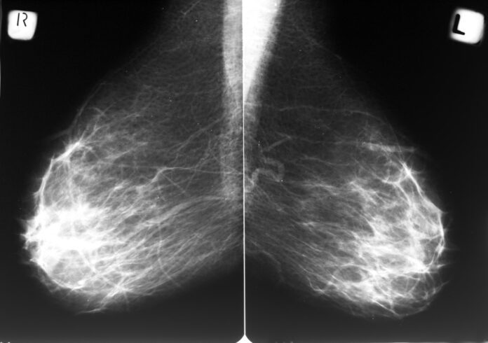
Most people who have Paget disease of the breast also have one or more tumors inside the same breast. In addition to ordering a nipple biopsy, the doctor should perform a clinical breast exam to check for lumps or other breast changes. As many as 50 percent of people who have Paget disease of the breast have a breast lump that can be felt in a clinical breast exam. The doctor may order additional diagnostic tests, such as a diagnostic mammogram, an ultrasound exam, or a magnetic resonance imaging scan to look for possible tumors.
Source
https://rarediseases.org/rare-diseases/pagets-disease-of-the-breast/
https://www.cancer.gov/types/breast/paget-breast-fact-sheet
https://www.cedars-sinai.org/health-library/diseases-and-conditions/p/pagets-disease-of-the-breast.html


